NEONATOLOGY ON THE WEB
Premature and Congenitally Diseased Infants
by Julius H. Hess, M.D.
Chapter XIII
Diseases of the Nervous System
Mental and Nervous Disturbances
The frequency of mental disturbances and other phenomena on the
part of the central nervous system in premature infants has been
variously estimated. Finkelstein states that mental disturbances and
spastic phenomena are not more frequent in prematures than in
full-term infants, but Ylppö strongly contests this statement.
The attempt to express the frequency of permanent mental defects and
other cerebral disturbances in percentages is only rarely possible
before the end of the first year of life, with perhaps the exception
of the typical Mongolian idiot. Demonstrable mental defects, either
complete idiots or imbeciles, were found in 7.4 per cent of
Ylppö's cases.
Mental defects in premature infants are frequently accompanied by
other symptoms on the part of the central nervous system. The most
common are the spastic paraplegias and diplegias. These are present
in prematures with demonstrable mental defects in at least 75 per
cent of all cases. However, mental development may be complete in the
presence of spasticity of the extremities dependent upon cerebral
irritation. In most instances this is secondary to intracranial
hemorrhage. Paraplegia or diplegia was present in 3.1 per cent of all
Ylppö's cases. These figures would certainly be much higher, had
all the prematures remained alive, since most of the infants
suffering from injury to the brain die very early. The cerebral
affections occur the more frequently, the smaller the infant at
birth.
In our experience mental disturbances and defects on the part of
the central nervous system have been confined largely to those
infants who survived from among the class of so-called
weaklings. These are the infants who have suffered from
intra-uterine disease or congenital malformations, traumata at birth,
or postpartum dietetic errors and infection. Among the more mature
that are normal for their fetal age the prognosis for a full mental
development is good.
Treatment. -- In the postmortem examination of infants
dying of cerebral hemorrhage, Rodda [1] found over
50 per cent followed non-instrumental deliveries and many followed
normal and easy births. In these cases the blood was found slightly
or not at all coagulated. Cerebral hemorrhage was by far the most
frequent cause of death in the new born in his group of cases. In
many cases at postmortem, no torn veins were found in the cerebrum or
cerebellum to account for the hemorrhage, and multiple hemorrhages
were found in portions of the body where it was inconceivable that
they could be explained by trauma. Over 25 per cent of all infants
dying of cerebral hemorrhage showed this picture of multiple
hemorrhages. An analysis of cases reported in the literature deepened
the conclusion that these hemorrhages were due to factors other than
trauma. Further study led to the conclusion that there was a
disturbance in the coagulation time of the blood in the new born. It
was found that the average coagulation time in the new born was seven
minutes. In icterus, melena, jaundice, syphilis and non-traumatic
cerebral hemorrhage, the coagulation time of the blood was prolonged.
In melena it might be delayed to ninety minutes. The subcutaneous
injection of normal blood was effective in cases in which there was
delayed or slow bleeding.
The further treatment in those cases with a diagnosis of
intracranial hemorrhage is symptomatic and expectant. There is always
the possibility that there may be spontaneous cure. The infant must
be kept quiet and warm. For the motor hyperirritability and
convulsions narcotics may be employed, before all chloral hydrate
(0.12 to 0.5 gm per day per rectum), also bromides (0.25 to 1 gm. per
day) or calcium lactate (1 to 2 gm.) or calcium bromide (0.3 to 0.5
gm. per mouth) per day.
Where the infants do not swallow well, feedings must be given per
catheter or subcutaneous infusions must be used for emergency.
Lumbar puncture, although primarily a diagnostic measure, may have
a beneficial therapeutic action. It is of diagnostic value when the
hemorrhage is below the tentorium.
In the full term, cranial decompression when employed early has
yielded favorable results, however little can be expected from such
surgical interference in the premature.
Schulze's swingings and other violent measures for artificial
respiration are distinctly contraindicated in the treatment of
asphyxia in the premature.
For the paraplegias and diplegias, corrective measures should be
undertaken early in order to prevent marked deformities. Massage and
active and passive movements should be practised regularly beginning
to advantage in the first year.
Muscle training in walking, climbing, and other activities should
be instituted under the supervision of a trained assistant.
Orthopaedic appliances are frequently indicated.
Surgical procedures may be necessary later.
Another group is made up of premature infants with more or
less serious mental defects in whom typical epilepsy gradually
develops, often of the Jacksonian type. It is very difficult and
often impossible to make a differential diagnosis between epilepsy
and spasmophilia in the first attacks, and especially in those cases
where the convulsions appear very early. Fortunately, as a general
rule, the epileptic convulsions do not occur in the first year of
life in prematures, while on the other hand electric
hyperirritability and spasmophilic convulsions are quite frequent in
this period of life. This makes the differential diagnosis somewhat
easier.
On the other hand, however, in connection with febrile diseases of
later life, convulsions occur very frequently in premature infants.
Only the further course of the disease will show whether the
convulsions are of epileptic or spasmophilic nature.
Hydrocephalus; Megacephalus
True congenital hydrocephalus is usually of the internal type with
enlarged ventricles. The external form is very rare. Megacephalus
must be differentiated from hydrocephalus, the two often being
confused in the premature, as previously mentioned in the discussion
of Pathology and Rachitis. Internal hydrocephalus results from a
transudation or exudation. Obstruction to the outflow may be the
cause as in the case of intracranial hemorrhage or cerebellar cysts.
However, most of the cases are probably due to an intra-uterine
serous meningitis or meningoencephalitis of unknown origin. Syphilis
is frequently the cause of congenital hydrocephalus.
The inflammatory process bringing about hydrocephalus may be at
end by the time of completion of pregnancy, but usually persists
thereafter. Most of the infants show enlargement of the head soon
after birth or the enlargement becomes apparent at a later period.
When the process begins early, intra-uterine, it may bring about a
marked retardation in brain development. The head need not
necessarily be enlarged; indeed the head may be small as in a
microcephalic. The brain in these cases is really a large cyst. Often
the skull is enlarged at birth, and it may hinder labor to such an
extent that perforation or puncture of the head becomes necessary.
When the head has the classic hydrocephalic configuration the
diagnosis is, of course, easy. In many instances there are also the
following symptoms at birth: Hypertonus and spasms of the muscles,
increased reflexes, convulsions, psychic disturbances and apathy.
Where the characteristic head is not seen and only slight
enlargement of the fontanelle areas is noted, the diagnosis is
difficult. Intracranial hemorrhage and meningitis must be ruled out.
Lumbar or ventricular puncture is of great assistance.
The prognosis is usually difficult to make early. The only early
therapeutic measure is lumbar or ventricular puncture with drawing
off of cerebrospinal fluid. Late surgical interference may be
indicated.
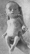
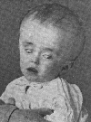
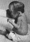
The term megacephalus is applied to conditions in which the
head develops out of proportion to the other body measurements and
length. It is characterized by an abnormally large head, with a
relatively larger brain. This condition is a characteristic finding
in a high percentage of infants prematurely born and is seen in
inverse proportion to the fetal age and birth weight. Rosenstern
[2], in a series of sixty-one prematures observed
over a period of at least three months, noted megacephalus in
forty-four. He concluded that the lower the birth weight of the
premature the more likely is megacephalus to develop.
Relation of Body Weight To Megacephalus
(Rosenstern)
|
Birth weight
|
No. cases
|
Present
|
Absent
|
|
Severe
|
Moderate
|
Mild
|
Total
|
|
to 1000 gm.
|
1
|
0
|
1
|
0
|
1
|
0
|
|
1001 to 1500 gm.
|
12
|
5
|
6
|
1
|
12
|
0
|
|
1501 to 2000 gm.
|
21
|
3
|
14
|
1
|
18
|
3
|
|
2001 to 2500 gm.
|
27
|
1
|
9
|
3
|
13
|
14
|
|
Total
|
61
|
|
|
|
44
|
17
|
It usually occurs before the age at which rachitic changes are
noted in the long bones and chest. However, as rickets occurs much
earlier in premature infants than in full-term infants, I do not
believe that we are at present in a position to dissociate these two
conditions. There is therefore great probability that the same
etiological factors underlying the development of megacephalus in the
first months may be the cause of rachitic manifestations in the bones
or other organs at later periods.
Time of Occurrence of Megacephalus (Rosenstern)
|
Age in months
|
Cases
|
|
1
|
9
|
|
2
|
11
|
|
3
|
11
|
|
4
|
0
|
|
5
|
4
|
|
6
|
0
|
|
7
|
1
|
|
8
|
1
|
|
9-11
|
0
|
|
12
|
1
|
It is most frequently seen during the second and third months of
life and reaches its maximum between the sixth and eighth months. It
then gradually becomes less manifest. There is usually an increased
spinal fluid pressure in which it resembles hydrocephalus. The brain,
on section, is found to be abnormally large but in true cases there
is a complete absence of hydrocephalus.
Associated with the large skull and wide-open fontanelles and
sutures, exophthalmos is frequently seen. The latter probably results
from the lack of skull capacity, the eyes being protruded, with
prominent cornea and, not infrequently, dilated pupils. Further
characteristics of the head are a broad face and mouth, nose and eyes
which appear closely set together: the nose is stumpy and small and
rises but little above the face; the tongue is often large and
protruded.
Encephalitis
The subject of encephalitis of the premature and full-term new
born is still very much in the dark. The etiology is obscure and a
clinical picture for the encephalitic processes has not yet been
described.
Encephalitis interstitialis congenita was described by Virchow
[3], with changes in the medullary substance of the
cerebrum, as a diffuse infiltration with fatty infiltrative cells.
Later, other observers declared that this was not pathological
(Jastrowitz [4], Limbeck [5]).
Brain defects (porencephaly) have been linked with congenital
encephalitis. Septic encephalitis is either a metastatic condition or
a meningo-encephalitis, the difference between the two being almost
impossible to define. The medullary substance shows clumps of
bacteria and leukocytes, and later there appears a suppurative
inflammation on the brain substance. Not infrequently in prematures
the meningo-encephalitis is a distinctly luetic process (see chapter
on Syphilis in Prematures).
Meningitis
The meningitic processes are as little understood as the
encephalitic. They may be acute or chronic. Serous meningitis which
is not well understood is supposed to be intimately related to
congenital hydrocephalus. Pachymeningitis hemorrhagica interna seems
to be a luetic process entirely (see Syphilis of Prematures).
Purulent meningitis follows suppurative conditions in the middle
ear, accessory nasal sinuses or is metastatic. Sometimes one sees
typical meningeal symptoms as: Convulsions, rigidity of the neck,
hypertonus, protruding fontanelles. However, meningitis may be
present without any characteristic signs. The infants are flaccid,
exhausted, and dried out. The diagnosis is verified by lumbar
puncture. Fever is often absent or only present terminally.
The prognosis is absolutely poor. Death usually follows in
twenty-four hours, but some linger eight to fourteen days. The
inception of the process is difficult to fix because of the
uncertainty of the symptoms.
Sinus thrombosis following middle-ear infections or phlebitis
after navel infections sometimes are responsible for the meningitis.
Epidemic cerebrospinal meningitis is not an uncommon complication
in premature infants during the first year. A spinal puncture should
be made in every case showing marked evidence of cerebral irritation.
In positive cases serum should first be administered intravenously
through the longitudinal sinus, because of the tendency to
generalization of the infection in this class of infants. Intraspinal
administration of serum must always be made by the gravity method
after withdrawal of as much fluid as is to be administered.
Finally we find among the prematures a number of idiots that have
to be classified as "degenerative idiots." These are the infants that
at birth already show stigmata of Mongolism or other malformations.
These children are prematurely born with special frequency, and it
follows therefore that a considerable number of children with
Mongolian idiocy are prematures. After all, it is a known fact that
children with various congenital malformations, be they congenital
bone diseases, bone anomalies, congenital heart disease,
malformations of the brain or spinal cord, etc., are born in an
immature condition. This circumstance, as previously mentioned, is
the reason that prematures have been very generally but erroneously
regarded as congenitally inferior.
Spasmophilic Convulsions
With reference to spasmophilic convulsions we must not regard them
as purely functional convulsions; on the contrary the readiness with
which they occur and their frequency in premature infants speaks very
strongly for organic lesions, probably most frequently among these
being cerebral hemorrhage occurring during labor. Naturally certain
extra-uterine noxae, as anemia and rachitis, are of importance as
determining factors that make the spasmophilic disturbances manifest.
Numerous roentgenological examinations of the long bones of premature
infants have shown that the rachitic changes are not confined to the
skull, but that the other bones are also early affected, as early as
the second and third months of life.
Footnotes
[1] Am. Jour. Dis. Child., 1920, 19, 268.
[2] Rosenstern, J.: Ztschr. f. Kinderh., 1922,
22, 129.
[3] Virchow's Arch., 1867, 38, 129.
[4] Arch. f. Psych., 1870, vol. 2 and 1871,
vol. 3.
[5] Prague Ztschr. f. Heilk., 1885, 7, 87.
|

|
Fig. 162. Megacephalus. Baby P. H. at four months.
|
|

|
Fig. 163. Baby P. H. at six months.
|
|

|
Fig. 164. Weight and food curves and calor intake.
Estimated fetal age, 32 weeks. One of triplets. Second died
shortly after birth. Third born macerated. Born April 1,
admitted April 1. Discharged, October 12; age one hundred
and ninety-three days. Birth weight, 840 gm; lowest weight,
645 gm.; doubled birth weight in one hudred and fifty-three
days; tripled lowest weight in one hundred and seventy-five
days. (From service of Dr. L. E. Frankethal, Michael Reese
Hospital.)
|
|

|
Fig. 165. Hydrocephalus. First signs when infant was four
weeks old.
|
|

|
Fig. 166. Oxycephalus (Tower skull). Usually associated
with other congenital defects and stigmata of degeneration.
The skull is dome shape with bulging temporal regions. The
deformity was present at birth. It is generally associated
with exophthalmos, propotosis and frequently with other
ocular abnormalities. Some children are mentally normal.
Others subnormal.
|
Return to the Hess Contents Page
Created 5/27/97 / Last modified 5/28/97
Copyright © 1998 Neonatology on the Web / webmaster@neonatology.net


